What’s the Difference Between a CT Scan vs. MRI?
Updated: Mar. 08, 2022
CT scans and MRIs are common, noninvasive medical imaging techniques used to see what's happening inside the body. Learn the differences.
CT scan vs. MRI: What’s the difference?
When doctors need to know what’s happening in the body—without slicing you open—they turn to imaging tests. These medical marvels make it possible to get a good visual on what’s happening in your body. And that can lead to a diagnosis and treatment.
There’s a good chance you’ve had at least one type of imaging before: an X-ray, which takes two-dimensional pictures and is often used to see the bones. You probably remember getting X-rays at the dentist.
But if you’ve ever been injured or had unexplained medical symptoms, you might’ve undergone two other types of imaging tests: a computed tomography (CT) scan or a magnetic resonance imaging (MRI) scan.
These are among the two most commonly used non-invasive imaging methods that can help doctors give you a more accurate diagnosis.
Although both a CT scan and an MRI are considered to be low risk, choosing between the two is done on a case-by-case basis.
Here’s everything you need to know about the differences between a CT scan and an MRI, including when doctors use them and the pros and cons of each.
What is a CT scan?
A CT scan is a computerized X-ray imaging procedure in which a narrow beam of X-rays is aimed at a patient and quickly rotated around the body.
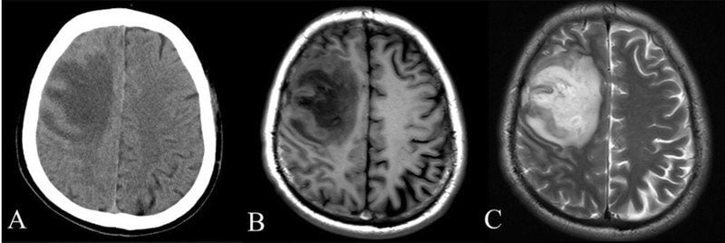
The X-rays are absorbed by detectors and processed by the machine’s computer to create cross-sectional images, or “slices,” of the body. More detailed than a traditional X-ray, these slices are called tomographic images.
The cross-sectional images can also be digitally stacked together to form a three-dimensional image for easier identification and location of basic body structures and possible tumors or abnormalities.
“CT scans revolutionized medicine and how we look inside the body and soft tissues,” says Claudia Kirsch, MD, division chief of neuroradiology at Northwell Health in Manhasset, New York. “They allowed detailed visualization of the brain, vessels, and soft tissues compared to standard X-rays.”
How does a CT scan work?
Unlike conventional X-rays, CT scans use a motorized X-ray source that rotates around the circular opening of the machine’s gantry, a doughnut-shaped structure.
A person lies on a bed that moves through the gantry while the X-ray tube rotates around the patient, shooting narrow beams of X-rays through the body.
Instead of film, the machine uses digital X-ray detectors that are located opposite the X-ray source. As X-rays leave the patient, they’re picked up by the detectors and transmitted to a computer. (Hence the “computed” in the name.)
Each time the X-ray source completes a full rotation around the body, the machine uses a mathematical technique to construct a three-dimensional image of the anatomy.
Once a full slice is completed, a 360-degree image is stored and the bed is moved forward incrementally into the gantry, where it completes another rotation.
The process continues until the technician has the number of slices needed to create detailed images of internal organs, blood vessels, and bones.
“Each scan is made up of hundreds, or maybe thousands, of images that are put together to construct whole pictures,” says Jonathan Goldin, MD, a diagnostic radiologist at UCLA Medical Center in Santa Monica, California.
(Don’t miss the doctors you should see at every age.)
How do doctors use CT scans?
Dr. Goldin says the purpose of a CT scan is to look for diseases or injuries based on a patient’s symptoms or risk factors.
The detailed pictures of organs, bones, and other tissue help diagnose abnormalities and can guide doctors to the right area during a biopsy or treatment procedure.
The goal of a radiologist is to put together visual information, combine it with clinical information, and suggest a diagnosis or other kinds of testing, says Adam Bernheim, MD, a cardiothoracic radiologist at Mount Sinai in New York.
“CT scans are readily available and are often performed in emergency settings because they can quickly rule out and rule in diseases,” says Dr. Bernheim, who is also an associate professor of diagnostic, molecular, and interventional radiology at the Icahn School of Medicine at Mount Sinai.
What are the downsides of a CT scan?
CT scans can be incredibly helpful in diagnosing injuries and diseases doctors can’t perceive with the eye or can’t pinpoint by taking your medical history and doing an evaluation.
That said, they’re not entirely risk-free. There are downsides for certain people.
Risk of allergic reaction
Some CT scans require dye for contrast. It’s provided before the procedure to highlight certain areas of anatomy on the scan.
Rarely, some people have an allergic reaction to the dye. The reaction depends on how severe your allergy is, but you might get hives, wheezing or shortness of breath, swelling, low blood pressure, a racing heart, and swelling in your throat.
If you know you’re allergic, you can take medication to mitigate the risk. The trick, of course, is knowing ahead of time.
Dye can be injected in several ways. Sometimes people drink it before a scan. Sometimes it’s injected through an IV (intravenous) in the hand or forearm. And sometimes, though rarely, it is given through the rectum using an enema.
Given as an IV, contrast may cause a slight burning feeling, a metallic taste in the mouth, or a warm flush in the body.
Most contrast injected into the vein contains iodine, so if a patient is allergic to iodine, it may cause nausea, vomiting, sneezing, itching, or hives.
Risk for people with diabetes
Diabetes raises the risk for a few different side effects, thanks to the contrast dye.
A person with diabetes may have to stop taking metformin before getting contrast because the medication has been linked to rare but serious side effects when combined with a contrast agent.
Another rare side effect is something called contrast-induced nephropathy (CIN), a decline in kidney function.
The reaction isn’t common and can be reversed, but having diabetes ups your risk. And having both diabetes and chronic kidney disease takes your risk of CIN from about 2 percent to 20 to 5o percent, according to the National Kidney Foundation.
Finally, if you have diabetes (or hypoglycemia), keep in mind that if contrast is used, you may not be able to eat or drink for four to six hours before the test. You’ll want to schedule the test accordingly so your blood sugar doesn’t drop too low.
Risk for people with kidney disease
A kidney problem may be a reason to stay away from contrast, as it worsens function.
Not only can contrast cause CIN in a small group of people, but the National Kidney Foundation notes that risk for the rare condition jumps from 2 percent to 30 to 40 percent if you have chronic kidney disease.
Kidneys remove iodine from the body, so if you have kidney problems, you may need extra fluid after the scan to flush the iodine out.
Risk for children and pregnant women
CT scans expose patients to radiation, though the risk has to be weighed against the benefits.
“The CT scan is an incredibly powerful tool that can make diagnoses and provide valuable information that should only be used when benefits outweigh the risks,” says Dr. Bernheim.
But while the benefit usually outweighs this risk, that’s not always the case for children. That’s why abdominal CT scans during pregnancy are considered particularly risky.
Children are more sensitive to ionizing radiation, and because exposure at such a young age is more likely to increase their risk for developing a future cancer.
Machine settings, however, can be adjusted for children to minimize radiation dose.
Risk of false positives
“One of the drawbacks of CT scans is the discovery of incidental false positives that can cause great anxiety,” says Dr. Bernheim.
That is, a scan might show a tumor. You might get (understandably) alarmed only to find out later that it was a false positive and you’re in the clear. (Note: There is also a risk of false positives with MRIs as well.)

What is an MRI?
Like the CT scan, the discovery of the MRI was a milestone in medicine.
An MRI is a medical imaging technique that uses a strong magnet to create detailed pictures of organs and tissues in the body.
The machine looks like a large tube with a bed running down the middle. A patient lies on the bed, and it slowly moves into the tube for scanning.
“An MRI machine is incredibly powerful and is often used in conjunction with a CT scan to get a more detailed look at a problem or condition,” says Dr. Bernheim. “It allows doctors, scientists, and researchers to examine the inside of the human body in high detail using another noninvasive tool.”
How does an MRI work?
The human body is composed of water molecules made of hydrogen and oxygen atoms. At the center of each atom is a small particle called a proton, which is sensitive to magnetic fields.
Water molecules in the body are randomly arranged, but when a patient enters an MRI scanner, an initial magnetic pulse causes those water molecules to align in one direction, either north or south.
An additional magnetic pulse is then quickly turned on and off, causing each hydrogen atom to change alignment. This repeats itself in a series of switches.
Electricity passes through gradient coils that cause them to vibrate and create a magnetic field. The process causes a knocking sound inside the scanner. (It’s loud. You’ll hear it even with earplugs or headphones.)
During the scan, which can take anywhere from 20 to 60 minutes, depending on the body part being scanned, you must stay still because movement will distort the scanner and create blurry images.
How do doctors use MRIs?
The MRI machine is a very expensive, sophisticated piece of equipment, and it can provide a more detailed picture of what’s going on in your body.
“An MRI can provide a doctor with more information about something seen by a CT scan and is particularly good at characterizing soft tissue,” says Dr. Bernheim.
It’s often used when an X-ray or CT scan doesn’t provide enough information, and a more detailed look is needed. For example, notes Dr. Bernheim, a CT scan may be used for a quick look at the brain and if necessary, an MRI scan will take a closer look.
Sometimes your doctor might jump straight to an MRI.
“While a CT scan measures density, an MRI can provide a complementary and detailed look at anatomy and pathology,” says Dr. Bernheim.
What are the downsides of an MRI?
There are some big upsides to an MRI: Little preparation is required, and unlike the CT scan, there is no radiation involved. However, they are a few drawbacks.
People with medical devices or implants can’t get an MRI
The MRI machine is basically one huge magnet, so no metal object can be anywhere in the room. That means you’ll have to remove all jewelry, watches, and other metals from you before you get the scan.
If you have metal in your body—say, shrapnel, a bullet, or medical devices like pacemakers or implants—you cannot have an MRI.
It’s not ideal if you’re claustrophobic
To have an MRI, you have to be in a relatively enclosed space. That’s usually well-tolerated, but if you’re very claustrophobic, you may need calming medication.
Open MRIs are available for certain body parts.
It’s loud—very loud
When inside the machine, the magnets create a loud, banging noise. Headphones or earplugs are often given to patients to dampen the noise.
You have to stay very still
Because movement can disrupt images, patients have to remain still, sometimes for as long as an hour or more. That’s usually doable, but expect to suddenly itch everywhere simply because you can’t move.
Pregnant women should avoid MRI dye injection
An IV of contrast dye is sometimes used with an MRI to improve visibility of a specific tissue and can cause rare side effects, says Dr. Bernheim.
The dye used for MRIs is different than that used for CT scans and should be avoided in pregnancy until the second and third trimesters.
Generally, though, MRIs are safe at a low measurement of magnetic strength. Sometimes, says Dr. Goldin, if scans take too long, body temperature may rise, but this is of little clinical concern in diagnosis.
It’s expensive
A good, top-of-the-line MRI machine costs an excess of $1 million, and the cost of a scan is much greater than the cost of a CT scan.
How that affects you will depend on your health insurance provider, so you may want to check first.
A radiologist’s expertise is vital to health outcomes
People are often aware that a good doctor can make the difference in diagnosing and treating injuries and diseases. What they don’t often realize is that the radiologist doing your scan is just as important.
Findings are dependent on who reads your CT or MRI images, says Dr. Goldin. The expertise and reputation of a radiologist is vital to the quality of diagnosis and treatment received.
“Radiology has become so important, and as the quality of imagery improves, and the more those images are used for diagnostic and treatment guidance, the more specialized the field of radiology becomes,” says Dr. Goldin. “Today there are specialties and subspecialties in radiology, and piecing together and reading images can determine a patient’s length and quality of life.”
Next, these are the DIY home-health tests that could save your life.


